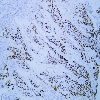
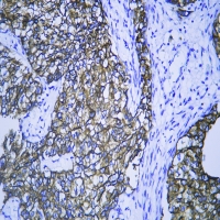
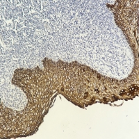
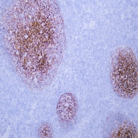
disease:Defects in INVS are the cause of nephronophthisis type 2 (NPHP2) [MIM:602088]; also known as infantile nephronophthisis. NPHP2 is an autosomal recessive disorder resulting in end-stage renal disease. It is characterized by early onset and rapid progression. Phenotypic manifestations include enlarged kidneys, chronic tubulo-interstitial nephritis, anemia, hyperkalemic metabolic acidosis. Some patients also display situs inversus. Pathologically, it differs from later-onset nephronophthisis by the absence of medullary cysts and thickened tubular basement membranes and by the presence of cortical microcysts.,domain:The D-box 1 (destruction box 1) mediates the interaction with APC2, and may act as a recognition signal for degradation via the ubiquitin-proteasome pathway.,function:Required for normal renal development and establishment of left-right axis. Probably acts as a molecular switch between different Wnt signaling pathways. Inhibits the canonical Wnt pathway by targeting cytoplasmic disheveled (DVL1) for degradation by the ubiquitin-proteasome. This suggests that it is required in renal development to oppose the repression of terminal differentiation of tubular epithelial cells by Wnt signaling.,PTM:May be ubiquitinated via its interaction with APC2.,sequence caution:Contaminating sequence. Potential poly-A sequence.,similarity:Contains 16 ANK repeats.,similarity:Contains 2 IQ domains.,subcellular location:Associates with several components of the cytoskeleton including ciliary, random and polarized microtubules. During mitosis, it is recruited to mitotic spindle. Frequently membrane-associated, membrane localization is dependent upon cell-cell contacts and is redistributed when cell adhesion is disrupted after incubation of the cell monolayer with low-calcium/EGTA medium.,subunit:Binds calmodulin via its IQ domains. Interacts with APC2. Interacts with alpha-, beta-, and gamma-catenin. Interacts with N-cadherin (CDH2). Interacts with microtubules (By similarity). Interacts with NPHP1. Interacts with DVL1, PRICKLE (PRICKLE1 or PRICKLE2) and Strabismus (VANGL1 or VANGL2).,tissue specificity:Widely expressed. Strongly expressed in the primary cilia of renal tubular cells.,
Cytoplasm . Cytoplasm, cytoskeleton . Cytoplasm, cytoskeleton, spindle . Membrane ; Peripheral membrane protein . Nucleus . Cell projection, cilium . Associates with several components of the cytoskeleton including ciliary, random and polarized microtubules. During mitosis, it is recruited to mitotic spindle. Frequently membrane-associated, membrane localization is dependent upon cell-cell contacts and is redistributed when cell adhesion is disrupted after incubation of the cell monolayer with low-calcium/EGTA medium.
Widely expressed. Strongly expressed in the primary cilia of renal tubular cells.





 下载说明书